What Are Organelles?
All cells are self-sustaining entities with different tasks divvied up to different components of the cell. These components are what we call cell organelles. Cell organelles perform important tasks to maintain normal cell functions including cell division. For Advanced Placement (AP) Biology there are two main eukaryotic cell types, the animal cell and the plant cell, which you need to understand well. A eukaryotic cell is a type of cell that possesses a distinct membrane-bound nucleus, whereas a prokaryotic cell does not have a nucleus.Here we will focus on eukaryotic cells.
For the purpose of this AP® Biology crash course, we will take a tour through plant and animal cells. We’ll look at which organelles they share, which ones they do not, why this is the case and the function of all these organelles. We will also touch up on how organelles are often strategically positioned.
Center of Operations: The Nucleus
We will start with the nucleus; both plant and animal cells have this organelle. The nucleus is the center of all cell functions. All but a few cell organelles rely on the nucleus for guidance. The nucleus is often located near the center of the cell. It is here that DNA is stored and RNA is synthesized, amongst other operations. Most of the cell’s ribosomal RNA synthesis activities take place in a cell organelle called the nucleolus, which is a small compartment of the nucleus. The nucleus as a whole is enclosed in a nuclear envelope, which is a double membrane that holds the content of the nucleus in place. This membrane extends onto the rough endoplasmic reticulum (ER), which we’ll touch upon next.
Manufacturing Organelle: The Endoplasmic Reticulum
For AP® Biology you will need to know the ER. The endoplasmic reticulum is a major manufacturing and transportation center of the cell with an intricate membrane system consisting of a network of tubules and sacs. This membrane system divides the internal sac space of the ER, the lumen, from the cytosol. In a nutshell, the endoplasmic reticulum is tasked with transporting materials within the cell.
The ER is divided into two types, the smooth endoplasmic reticulum (SER) and the rough endoplasmic reticulum (RER). The smooth ER is named for its relatively smooth exterior as comparedto the rough ER, which is covered in ribosomes on its cytoplasmic side. The SER carries outmetabolic processes including making lipids, phospholipids, and steroids. It is also charged with the removal of toxins, especially in liver cells.
The RER manufactures proteins and membrane parts. The ribosomes attached to the RER surface are vital to its protein synthesis function. Ribosomes can also be found floating in the cytoplasm and synthesizes proteins needed to run the cell. Ribosomes are complexes of RNA and proteins; they are not membrane-bound. Besides protein synthesis, the RER also performs post-translational modifications—modifying proteins after translation and preparing them for their respective functions. These modifications include processing and sorting secreted proteins.
The ER in plant cells has additional functions that you do not find in animal cells. It plays a central role in cell-to-cell communication in specialized cells. The ER is connected between plant cells through the plasmodesmata. Plant ER also stores proteins and contains additional enzymes and structural proteins that play a role in the biogenesis of oil bodies (lipid-containing structures) and the storage of lipids.
Processing and Sorting: The Golgi Apparatus
The Golgi apparatus, named for its discoverer Camillo Golgi, processes and sorts secreted proteins and membrane proteins from the RER. The Golgi apparatus is made of flattened, stacked membrane disks known as cisternae; stacks of cisternae are referred to as dictyosomes. The organelle has two distinct sides to it, one which receives materials, and the other which ships products out. Because the Golgi apparatus receives secreted molecules from the RER, it usually sits next to RER.
Even though the Golgi sits so close to the RER, the two organelles are not connected, and proteins still have to make their way from the RER to the Golgi. This is where transport vesicles come in; they are little membrane sacs that pinch of the ER membrane, travel to the ER, and deposit their contents in the Golgi apparatus by fusing with its membrane.
The proteins then move through the Golgi, getting modified along the way. Proteins leave the Golgi by budding off a bit of the Golgi membrane, using this membrane sac as a transport vesicle to the next organelle or for export from the through a process termed exocytosis. Essentially the Golgi apparatus, like the post office, is used for sorting molecules from the ER and ribosomes and shipping them to the rest of cell.
In plant cells, this cell organelle has an additional function that it does not need to perform in animal cells. The Golgi is a major biosynthetic organelle in plant cells and manufactures large amounts of polysaccharides that are required for cell wall synthesis.
We have covered the path ofRER manufactured products through the Golgi apparatus; now let us move on to what the Golgi does with molecules that need to be broken down. The Golgi can create other cell organelles that break down molecules just by budding off its membrane. Lysosomes are one such example.
Cellular Recycling Bins: Lysosomes
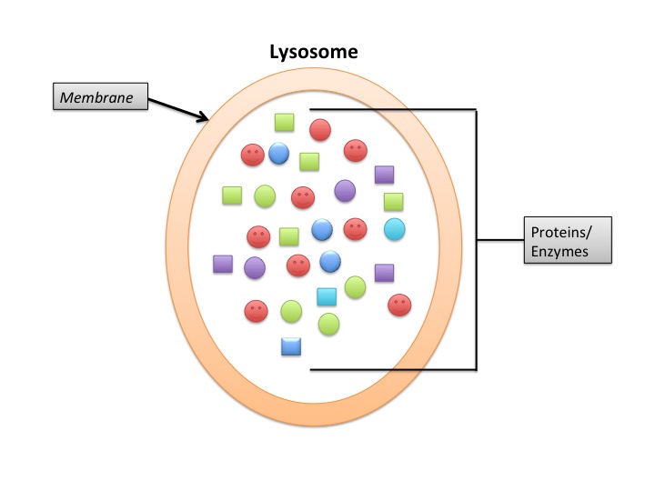
Lysosomes are produced in the Golgi and are membrane-enclosed sacs of hydrolytic enzymes that digest all major types of macromolecules. In short, worn-out materials floating around the cell are internalized and broken down by the lysosomes.
Think of lysosomes as recycle bins. If they were not functioning properly, the cells would be filled with access material they cannot handle. Diseases can develop from enzyme build-ups or leakage from the lysosome. Pompe’s disease is one of many lysosomal storage disorders (LSDs), all caused by something going pear-shape in the lysosome. In the case of Pompe’s disease, it results from the deficiency of carbohydrase (the enzyme responsible for the breakdown of glucagon) in the lysosome.
The recycling activities of the lysosome are referred to as autophagy, which means eating oneself—the cell breaks down some of its own components. Autophagy is the process of using hydrolytic enzymes in the lysosome to degrade old organelles. Autophagy is necessary because the cellengulfs external molecules through a process known as phagocytosis. Many of these molecules are large and insoluble–some as large as bacteria!—and lysosomes contain a variety of enzymes that break down these molecules into units as small as monomers. This is how old DNA and RNA can be broken down back into spare bases (Adenine, Guanine, Cytosine, Thymine, and Uracil) for re-use in DNA synthesis. Proteins and polypeptides are also broken down here by lysosomes.
Lysosomes are commonly found in animal cells but rarely ever in plant cells. This is because the central vacuole already performs the functions of lysosomes.
Central vacuoles are only found in plant cells and are tasked with the breaking down of molecules (we will revisit this cell organelle later).
Transportation within the Cell: Vesicles
Now that we have covered the recycling function of lysosomes let us talk about vesicles, the vehicles of the cellular shuttling system. These organelles are small compartments enclosed in a lipid bilayer membrane and are important for transporting molecules from one organelle to the next. Think of it this way; there is no point in dividing tasks amongst the cell organelles to achieve a common goal unless the cell has a transportation system to link all the organelles together.
Vesicles are filled with fluid they inherit from the parent organelle (e.g. Golgi) and are formed by pinching off of other organelles when materials need to be transported out of the cell or between organelles within the cell. Thus, vesicles form during exocytosis (secretion) or phagocytosis (uptake). When vesicles get to their destination, they can simply fuse with the membrane of the target organelle and deposit their content. Vesicles are also responsible for enzyme storage since they act as pocket reserves and can thus have a pH different from that of the cytosol. This is important because the vesicles can maintain conditions specific for the enzymes within without disrupting the rest of the cytosolic conditions.
There are many different types of vesicles. Secreted proteins are carried in secretory vesicles, which fuse with the membrane of the target organelle and release the proteins into the target organelle. There are also special vesicles called peroxisomes. Peroxisomes are tasked with detoxifying the cell by breaking down toxic molecules. They are also involved in biosynthesis by breaking down fatty acids into components used to make new fatty acids.
Peroxisomes look similar to lysosomes, but they are bigger in size. Peroxisomes come from the ER or other peroxisomes ; lysosomes, however, come from the Golgi apparatus. Peroxisomes are found near mitochondria and chloroplasts whereas lysosomes can be found anywhere in the cell. Lysosomes are involved more on the moderation side of the business by maintaining healthy amounts of certain molecules. For example, while glycogen is not normally toxic to the cell it is bad in large amounts, so lysosomes moderate concentrations of this polysaccharide. The peroxisome, on the other hand, is tasked with a detoxing function. Toxic substances such as ethanol can be broken down by peroxisomes to manageable bits such as water and molecular oxygen.
Storage Compartments: Vacuoles
While we are on the topic of fluid-filled sacs, we will move on to the next type of organelle: vacuoles. Vacuoles are also large membrane-enclosed, fluid-filled sacs. There are several types of vacuoles, mostly responsible for storage. Food vacuoles are created through phagocytosis and carry out intracellular digestion. Contractile vacuoles are found in freshwater protozoans; their main function is to remove excess water from the cell by taking up water from the cell and contracting to pump the water out. There are other types of vacuoles tasked with storage of waste and toxins.
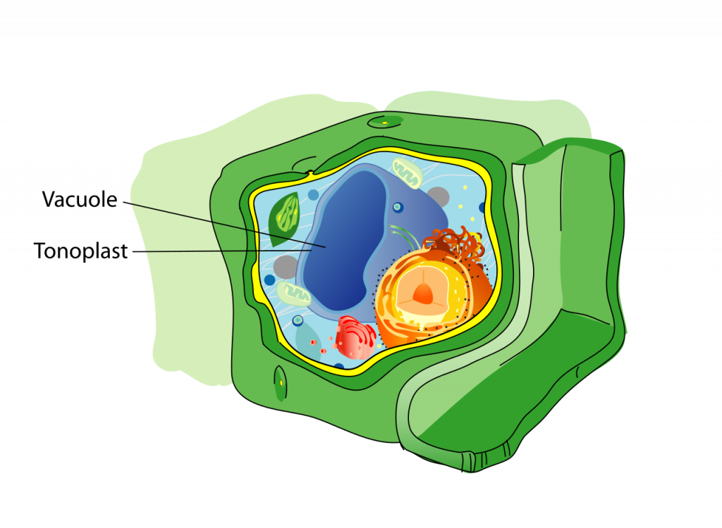
In most plant cells, you will encounter a central vacuole. The central vacuole forms as a result of smaller vacuoles from the Golgi and ER accumulating in the cell and joining to form the larger central vacuole. The functions of the central vacuole include storage of organic compounds or inorganic ions and sequestration of harmful cell by-products. The central vacuole can also help hold the cell’s shape as it enlarges. The central vacuole is specific to plant cells; it is not found in animal cells. The central vacuole plays a vital part in the maintenance of the cell’s water concentration in the face of changing environmental conditions.
By now you may be wondering what the difference is between vesicles and vacuoles. They are similar–both vesicles and vacuoles are membrane-bound and are tasked with storage and transport. However, vacuoles are usually larger than vesicles. Also, the membranes on vesicles are made such that they can fuse with membrane systems in the cell when they deposit their contents. This ability to fuse with membranes makes sense since many vesicles are formed by budding off the Golgi membrane. The membranes of vacuoles do not fuse with other cell or organelle membranes.
Organelles from Elsewhere: Mitochondria and Chloroplasts
Some organelles are hypothesized to have been independent entities taken in by eukaryotic cells by phagocytosis. These organelles are called plastids and include organelles such as mitochondria and chloroplasts, which are not fully dependent on the nucleus. Although they have since donated some of their genes to the nuclear genome, they still have their own genome that is unique from the nuclear genomes. However, compared to those of free-living bacteria such as cyanobacteria, these genomes are significantly reduced. This is to say that, the chloroplasts of free-living bacteria are far more complex with more genes than those of the plant-contained chloroplast.
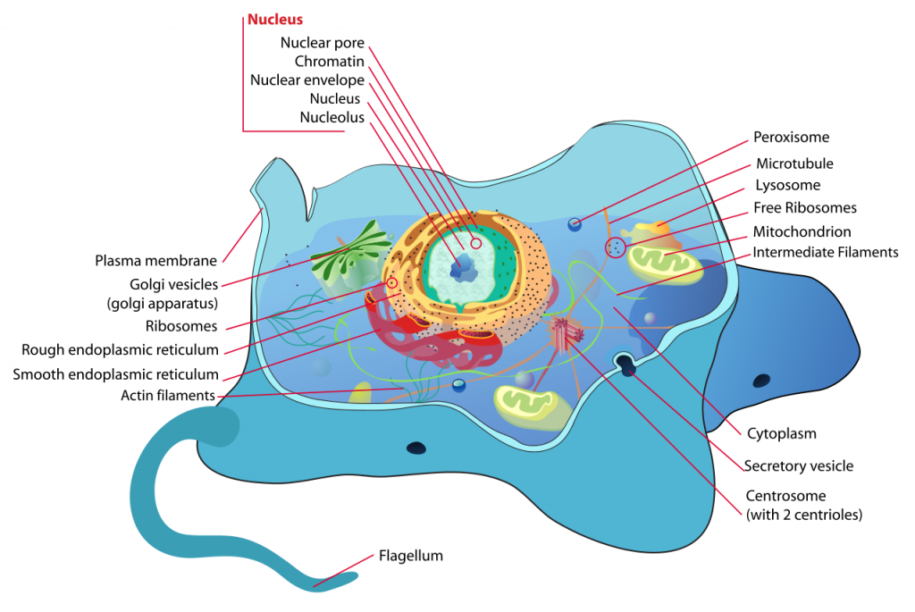
Mitochondria, the powerhouses of the cell, are found in almost all eukaryotic cells. The mitochondrion is enclosed by a double membrane. It converts energy from the surroundings into forms of energy accessible to the cell, such as ATP. The mitochondrion is encapsulated in two of its membranes, has ribosomes and DNA to synthesize proteins. The outer mitochondrial membrane is smooth and permeable to small solutes, but can easily block macromolecules. The inner membrane contains enzymes essential for cellular respiration. As you may have noticed, the mitochondrion is a fully functioning entity with its own machinery. It also has its own DNA, which is maternally inherited.
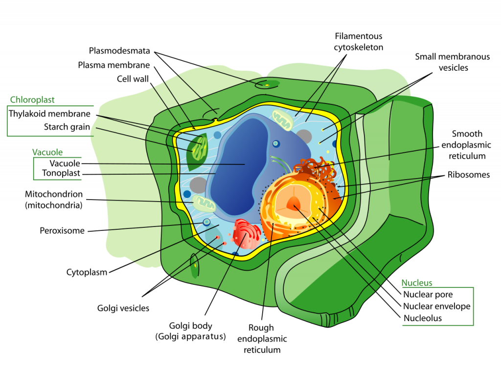
Chloroplasts are only found in plant cells and photosynthesizing bacteria such as cyanobacteria. They also have outer and inner membranes. The chloroplast consists of two main regions, the stroma and the thylakoid space (the space between the thylakoid membranes). The stroma is the fluid-filled space between the outer and inner membranes. In the thylakoid space, there is a stack of inner membranes called the thylakoid membranes where photosynthetic pigments such as chlorophyll are found. As you might have guessed, the chloroplast is the center of photosynthesis. Understanding this organelle is essential for AP® Biology since it also helps with your understanding of photosynthesis.
Other types of plastids exist such as leucoplasts. Leucoplasts are colorless organelles where starch and oils are stored; these are found in plants such as potatoes.
Keeping It All Together: Cytoplasm, Cell Membrane, Cell Wall
All of these organelles are suspended in cytosol inside the plasma membrane. Everything contained within the cell, except the nucleus, is collectively called the cytoplasm. The cytosol is the fluid part of the cytoplasm that contains water, salts, and other chemicals, and is jelly-like in its consistency. The organelles are suspended in the cytosol. The cytosol provides cushioning for the organelles.
Finally, the plasma membrane surrounds the cell and controls transportation into and out of the cell. The plasma membrane is selectively permeable; some substances are allowed entry while others aren’t. It demarcates where one cell ends and where the next one begins. It is made of proteins and fats. We also call it the cell membrane. This cell organelle is found in both animal and plant cells.
Last but not least, there is a second type of layer that encloses cells: the cell wall. This organelle is only found in plant cells but not in animal cells. It may interest you to know that cell walls can also be found in fungal cells, but the composition of fungal cell walls is different than that of plant cell walls. Plant cell walls are made mainly from cellulose. Their main function is to maintain the shape of the cell and protects against mechanical stress. Moreover, the cell wall prevents excess water uptake that might result in the death of the cell
Both plant and animal cells have a cytoskeleton that provides additional structural support. The cytoskeleton is a network of fibers that stretch throughout the cytoplasm and provides an intricate framework for support and movement. The cytoskeleton also associates with specialized proteins involved in movement such as muscle contractions.
We understand that remembering all of this can seem a little overwhelming, so for this AP® Biology crash course here is a table summarizing the major organelles.
Table 1: A summary of the major organelles and their functions
| Cell membrane | “The gate keeper.” Controls what comes in and what goes out of the cell. |
| Cell wall | Only found in plants, basically the tougher version of the cell membrane. It is stricter in what it lets in and it is made of cellulose in plants. |
| Cytoplasm | Everything in the cell except the nucleus including the cytosol and other organelles. |
| Mitochondria | The powerhouse. Think ATP synthesis. |
| Lysosomes | The recycle bin. It breaks down old by-products and organelles into smaller, reusable particles. |
| Vacuole | Storage centers for food, water, etc. Can store anything, really. |
| Golgi apparatus (remember to always capitalize the “G”) | The packaging and distribution center of the cell. It modifies products of the ER and ships them off to their destinations. |
| Endoplasmic Reticulum | The tunnel system of the cell. |
| Ribosomes | They make proteins from the triplet code of DNA. Think Central Dogma; this is where the genetic code is translated to make proteins. |
| The nucleus | This is the brain of the cell. The DNA code is stored here. |
| Chloroplasts | They are food manufactures. Energy from the sun is captured here and converted into food, water and oxygen. |
Hopefully at this point you understand what each cell organelle is and what it does. For AP® Biology you also should be able to identify plant and animal cell organelles based on their appearance and function. You should also understand why some organelles are limited to only plant cells or only animal cells.
In this AP® Biology crash course we have gone over how well the cell divvies up tasks to different organelles and how the outputs of each cell organelle reach their targets. Cells achieve an impressive amount of work, and they do so by trusting their individual parts to achieve their tasks for the bigger scheme of things. Both animal and plant cells are enclosed in membranes, but plant cells have an additional support, the cell wall. They each have cell organelles such as plastids, vesicles, nuclei, cytoplasm, and lysosomes. Mitochondria are limited to animal cells while chloroplasts are limited to plant cells. Cell organelles are vital to the functioning of all cells. We hope that this short article on cell organelles helps with your understanding of your AP® Biology content.
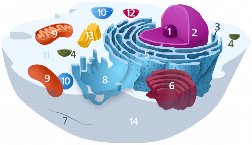
Test your knowledge
Can you label this diagram? Is this an animal or a plant cell?
Need help preparing for your AP® Biology exam?
Albert has hundreds of AP® Biology practice questions, free response, and full-length practice tests to try out.